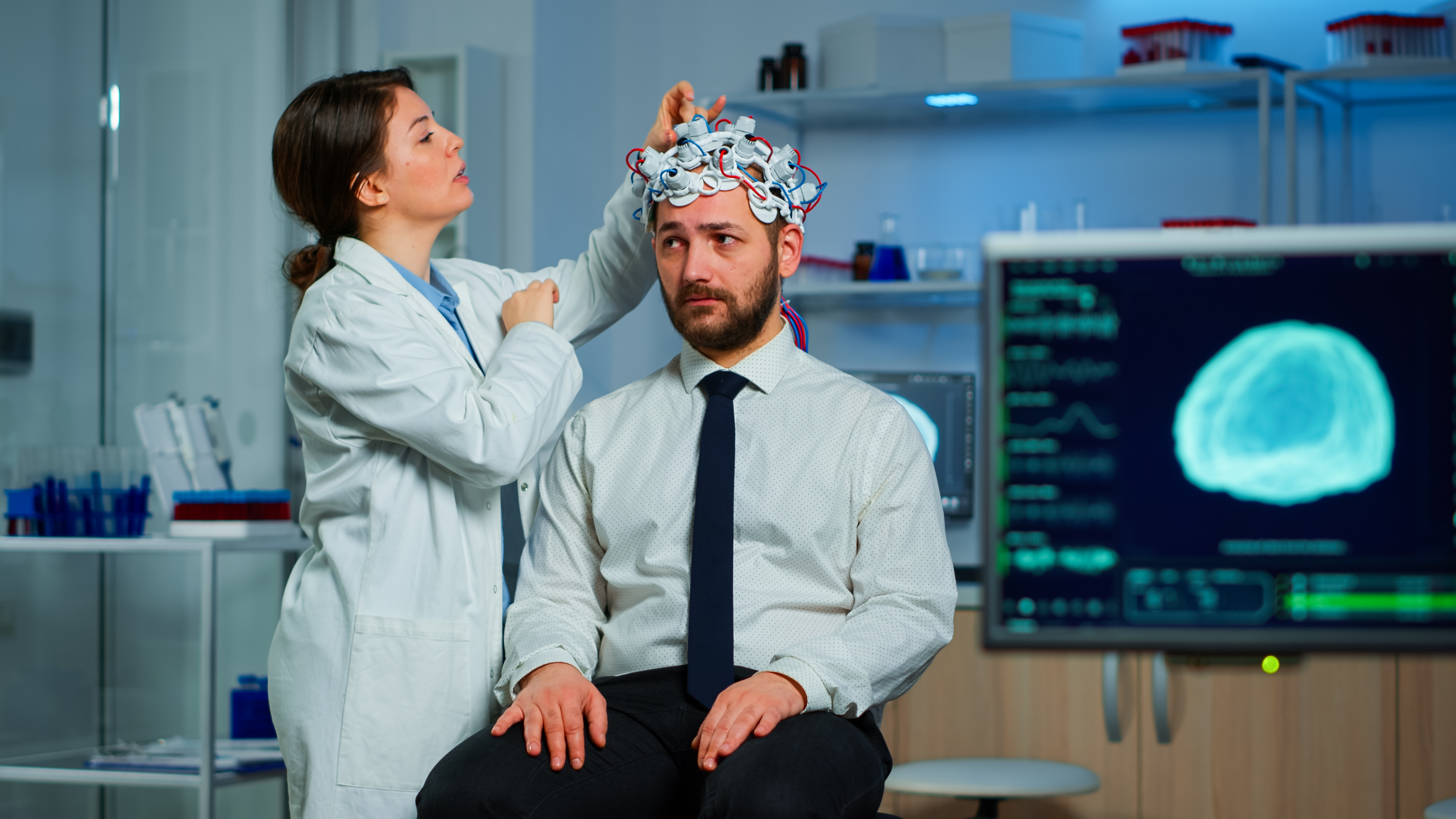
ELECTRO ENCEPHALOGRAPHY (EEG)
Electroencephalography (EEG) is a test that records electrical activity in the brain. It helps diagnose conditions like epilepsy, seizures, sleep disorders, and brain injuries. Small electrodes are placed on the scalp to detect brain wave patterns. The procedure is safe, non-invasive, and painless. EEG can be done while awake or during sleep, depending on the condition being assessed. It provides valuable insights into brain function and neurological health.
NCS (NERVE CONDUCTION STUDIES)
Nerve Conduction Studies (NCS) measure how quickly and efficiently nerves send electrical signals. They are used to diagnose nerve damage, pinched nerves, and neuromuscular disorders. Small electrodes are placed on the skin to stimulate nerves and record their responses. NCS is often performed alongside EMG (electromyography) for a complete nerve-muscle assessment. The test is generally well-tolerated, with minimal discomfort. Results help guide treatment for conditions like carpal tunnel syndrome or neuropathy.
CT (COMPUTED TOMOGRAPHY)
Computed Tomography (CT) is an advanced imaging technique that uses X-rays to create detailed cross-sectional images of the body. It helps diagnose conditions affecting the brain, lungs, abdomen, bones, and more. CT scans are quick, painless, and highly accurate. Sometimes a contrast dye is used to enhance image clarity. The scan provides vital information for detecting tumors, bleeding, infections, or injuries. CT plays a crucial role in emergency and routine medical care.
MRI (MAGNETIC RESONANCE IMAGING)
Magnetic Resonance Imaging (MRI) uses powerful magnets and radio waves to produce detailed images of internal organs and tissues. It’s especially useful for examining the brain, spine, joints, and soft tissues. Unlike X-rays or CT scans, MRI does not use ionizing radiation. The procedure is safe, non-invasive, and typically takes 30 to 60 minutes. Patients may need to lie still inside a tunnel-like scanner during the process. MRI helps diagnose conditions like tumors, strokes, ligament injuries, and multiple sclerosis.
NEURO-ULTRASOUND
Neuro-Ultrasound is a safe, non-invasive imaging technique that uses high-frequency sound waves to visualize brain structures. It is predominantly used in neonatal care to assess intracranial conditions through the open fontanelles. This method allows for real-time imaging, making it ideal for immediate bedside evaluations. Neuro-Ultrasound effectively detects abnormalities such as intracranial hemorrhage, hydrocephalus, and developmental anomalies. Its radiation-free nature makes it a preferred option for repeated examinations in vulnerable populations. Emerging applications are also exploring its utility in adult neurological assessments using specialized techniques.
OPHTHALMOSCOPIC EXAMINATION
Ophthalmoscopic Examination is a diagnostic procedure used to inspect the inside of the eye, particularly the retina and optic nerve. It helps detect signs of eye diseases, hypertension, diabetes, and neurological conditions. A handheld device called an ophthalmoscope shines light into the eye to view internal structures. The exam can be direct (simple handheld) or indirect (with a headlamp and lens for a wider view). It is quick, painless, and typically performed during routine eye or neurological exams. Changes in the retina may reveal early signs of systemic or neurological disorders.